
|
Features
|
Applications
|
Optical coherence tomography (OCT) is a rapidly developing optical imaging method that uses light to obtain images with high resolution (10 - 100 times greater than ultrasound) to depths of 1-3 mm in biological tissues and other materials. Applications include research in biology and medicine and imaging of diseased tissues in situ. OCT also has potential for non-destructive testing of specialized materials such as thermal barrier coatings used on jet engine turbines.
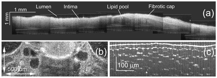
Examples of images obtained with spectral domain OCT at different center wavelengths and resolution. (a) atherosclerotic plaque (b) developing otic structures in an X.lavis tadpole (c) cellular structure in onion.
A proprietary, photonic-crystal fiber light source combined with spectral domain optical coherence tomography provides a versatile, spectrally agile instrument. The available spectral bandwidth is greater than needed even for 1 micron resolution in the near-infrared. This allows the user to select important optical parameters, such as the center wavelength and resolution, to match imaging requirements to specific applications. The system design allows simple, user-friendly operation, putting the focus on your research, not the instrument.
A limiting factor of all Fourier domain OCT methods is the complex conjugate
ambiguity due to Fourier transform of real-valued data; the image is symmetric
with respect to the zero plane of the interferometer. Only half of the imaging
depth range is useful in practice to avoid overlapping “mirror images.”
Resolving the complex conjugate ambiguity doubles the available imaging depth
range by allowing the interferometer zero plane to be within the sample. Modulating
the phase of the OCT light source separates the real and imaginary components
of the OCT return signal into components at the modulation frequency and at
twice the modulation frequency. More about this approach is in ref 1 and in
this poster presented at SPIE Photonics West/Bios 2010: High-Speed
Full-Range Imaging with Harmonic Detection Swept-Source Optical Coherence
Tomography.
Some of our recent publications in this area include
- "Resolving the complex conjugate ambiguity in Fourier-domain OCT by harmonic lock-in detection of the spectral interferogram," A. B. Vakhtin, K. A. Peterson, and D. J. Kane, Optics Letters 31, 1271-1273 (2006). Available at http://www.opticsinfobase.org,
- "A common path interferometer for frequency domain optical coherence tomography," A. B. Vakhtin, D. J. Kane, W. R. Wood, and K. A. Peterson, Applied Optics 42, No. 34 6953-6958 (2003).
- "Differential spectral interferometry: an imaging technique for biomedical applications," A. B. Vakhtin, K. A. Peterson, W. R. Wood, and D. J. Kane, Optics Letters, 28, No. 15, 1332-1334 (2003).
For more information, contact
Kris Peterson.
Contact Information
e-mail info@swsciences.com